Heart structure and the cardiac cycle
Select Lesson
Exam Board
Select an option
Practical skills
Control systems
Origins of genetic variation
Modern genetics
Microbiology and pathogens
Energy for biological processes
Exchange and transport
Classification and biodiversity
Cells, viruses and reproduction of living things
Biological molecules
Explainer Video

Summary
Heart structure and the cardiac cycle
In a nutshell
The heart is a vital organ that keeps organisms alive by pumping blood around the body. The heart is specialised to carry out its function and in this summary you will learn about the components of the heart, the sequence of events that happen during a heartbeat and how to interpret data.
The heart
The heart is made up of two main pump systems. The right side of the heart pumps deoxygenated blood to the lungs and the left side of the heart pumps oxygenated blood around the body. The structure of the heart is shown below.

| 1. | Superior vena cava | The superior vena cava transports deoxygenated blood from the head, neck, arms and chest to the right atrium. |
| 2. | Aorta | The aorta is connected to the left ventricle and it carries oxygenated blood to the rest of the body. |
| 3. | Pulmonary artery | The pulmonary artery carries deoxygenated blood away from the heart. |
| 4. | Right atrium | The right atrium is one of the four chambers of the heart. It receives deoxygenated blood from the body. |
| 5. | Right semi-lunar valve | The right semi-lunar valve is located between the right ventricle and the pulmonary artery. It prevents blood flowing backwards from the pulmonary artery into the right ventricle. |
| 6. | Right atrioventricular valve (tricuspid) | The tricuspid atrioventricular (AV) valve is located between the right atrium and the right ventricle. It has three flaps and stops blood flowing back from the right ventricle into the right atrium. |
| 7. | Right ventricle | The right ventricle is one of the four chambers of the heart. It receives deoxygenated blood from the right atrium and pumps it to the pulmonary artery. |
| 8. | Inferior vena cava | The inferior vena cava transports deoxygenated blood from lower parts of the body to the right atrium. |
| 9. | Pulmonary vein | The pulmonary vein carries oxygenated blood from the lungs to the heart. |
| 10. | Left atrium | The left atrium is one of the four chambers of the heart. It receives oxygenated blood from the lungs. |
| 11. | Left atrioventricular valve (biscuspid) | The bicuspid atrioventricular (AV) valve is located between the left atrium and the left ventricle. It has two flaps and stops blood flowing back from the left ventricle into the left atrium. |
| 12. | Cardiac muscle | The cardiac muscle is thick and helps the heart to pump blood around the body. |
| 13. | Left semi-lunar valve | The left semi-lunar valve is located between the left ventricle and the aorta. It prevents blood flowing backwards from the aorta into the left ventricle. |
| 14. | Left ventricle | The left ventricle is one of the four chambers of the heart. It pumps oxygenated blood into the aorta. |
Adaptations of the heart
The heart is a very important organ that keeps organisms alive. Therefore, it must be adapted to carry out its function efficiently. Some of these adaptations are explained below.
Adaptation | Explanation |
| Thick walls in the left ventricle | The left ventricle pumps blood around the body therefore it must contract more forcefully than the right ventricle which is only pumping blood to the lungs. The thicker walls of the left ventricle facilitate this. |
| Thicker ventricular walls than atrial walls | The ventricles have to pump blood out of the heart through the arteries. This is further than the atria which only need to pump the blood into the ventricles. Therefore, the ventricles will have thicker walls. |
| Atrioventricular (AV) valves | The AV valves stop blood flowing from the ventricles into the atria when the ventricles contract. |
| Semi-lunar valves | The semi-lunar valves stop blood flowing back into the heart from the arteries after the ventricles contract. |
| Cords | The cords attach the AV valves to the ventricles. This provides support and ensures they only open one way. |
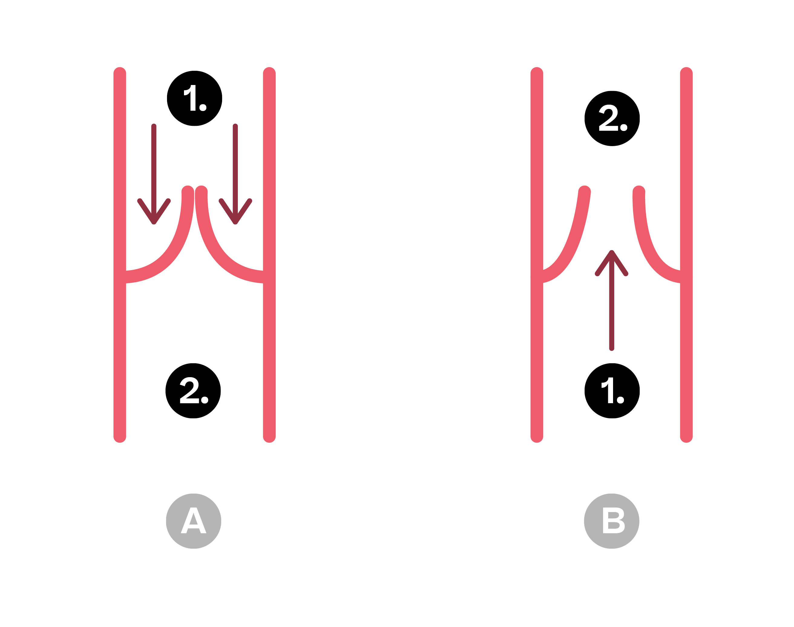
Valves
The cardiac cycle
Definition
The cardiac cycle is the sequence of events that takes place in one heartbeat. It is the sequence of contraction (heart empties) and relaxation (heart fills). Pressure and volume changes are experienced during the cardiac cycle. These are detailed below.

| | Phase | Explanation |
| 1. | Atrial systole | Blood under low pressure fill the atria through the pulmonary vein and vena cava. Increasing pressure inside the atria cause them to contract. The blood is forced through the atrioventricular valves into the ventricles. Note: The semi-lunar valves are closed. |
| 2. | Ventricular systole | The atria relax and after a slight delay, the ventricles contract. The pressure in the ventricles increases and blood is forced upwards, closing the atrioventricular valves. Blood is forced into the aorta and pulmonary artery, opening the semilunar valves. |
| 3. | Diastole | Both the atria and ventricles relax. Blood flows into the atria from the veins. Elastic recoil of the atrial walls generates a low pressure in the atria which helps to draw blood into the heart. Due to gravity, blood in the arteries flows down towards the ventricles. This closes the semilunar valves. |
Data interpretation
During the cardiac cycle, there are various pressure and volume changes that occur. You need to be able to interpret data regarding these changes. An interpretation of a graph can be shown below.

| 1. | Pressure increases due to the contraction of the atria. |
| 2. | Pressure decreases as the atria relax. |
| 3. | Pressure increases as the atria begin to fill. |
| 4. | Pressure drops a bit as some blood flows into the ventricles. |
| 5. | Pressure increases as the atria continue to fill. |
| 6. | Pressure increases by a small amount as they fill passively. |
| 7. | Pressure increases due to contraction of the ventricles. |
| 8. | Pressure decreases as the ventricles relax. |
| 9. | Pressure increases as the ventricles start to fill. |
| 10. | Ventricles stretch whilst filling so their volume increases. |
| 11. | When relaxed, the ventricles expand and fill with blood. |
| 12. | The volume of the atria decreases as they contract. |
| 13. | When relaxed, the atria expand and fill with blood. |
| 14. | Some blood passes through the AV valves and enters the ventricles so the volume of the atria decreases. |
Create an account to read the summary
Exercises
Create an account to complete the exercises

FAQs - Frequently Asked Questions
What do the atrioventricular valves do?
The atrioventricular (AV) valves stop blood flowing from the ventricles into the atria when the ventricles contract.
What happens during ventricular systole?
During ventricular systole, the atria relax and after a slight delay, the ventricles contract. The pressure in the ventricles increases and blood is forced upwards, closing the atrioventricular valves. Blood is forced into the aorta and pulmonary artery, opening the semilunar valves.
What does the pulmonary vein do?
The pulmonary vein carries oxygenated blood from the lungs to the heart.
Beta