Heart Anatomy: Labeled Diagram, Structures, Function, and Blood Flow
Anatomy of the human heart made easy using labeled diagrams of the main cardiac structures, along with their function, blood flow through the heart, and a review with a quiz at the end to test your knowledge!
Save Time with a Video!
Save time by watching the video first, then supplement it with the lecture below!
Click below to view the EZmed video library. Subscribe to stay in the loop!
💯 Pass Your Classes & Exams!
Instant Access to Hundreds of Study Guides, PDF Lectures, & Flashcards!
Instant access to a members-only page of ALL the flashcards, study guides, and PDF lectures. Cancel anytime.
Download the Study Guide!
SAVE time studying and ACE the anatomy of the heart with the study guide below!

Anatomy of the Heart
Welcome to the anatomy of the heart made easy!
We will use labeled diagrams and pictures to learn the main cardiac structures and related vascular system.
In addition to reviewing the human heart anatomy, we will also discuss the function and order in which blood flows through the heart.
By the end of this post, you will have a strong understanding of the main cardiac structures including:
Atria
Ventricles
Tricuspid Valve
Mitral Valve
Pulmonary Valve
Aortic Valve
Superior and Inferior Vena Cava
Pulmonary Arteries and Veins
Aorta
You will also be provided with numerous memory tricks to help you remember the different structures of the heart!
Make sure to check out the blank model of the heart at the end of this post, and quiz yourself on the gross anatomy by labeling and matching the main cardiac structures!
By the end of this post, you will be able to label a drawing of the heart similar to the anterior view (frontal section) shown below.
Let’s get started!

Image: Diagram showing the anatomy of the heart with the main structures labeled.
Heart Chambers - Atria and Ventricles
As we walk through the main anatomical structures of the heart, here are some other EZmed references you may find useful to help bring it all together!
The Cardiac Cycle - Step-By-Step Guide of the Heart Cycle from Diastole to Systole
Conduction System of the Heart - Step-By-Step Guide of the Conduction System
Now let’s really get started!
Let’s first use the simplified cartoon diagram below to walk through the main anatomical features of the heart.
We will then apply those structure to the more realistic diagram of the heart above.
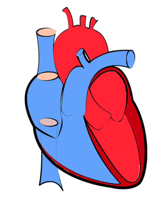
Image: Cartoon diagram of the heart we will use to review the main cardiac structures and anatomy.
Chambers of the Heart
Let's begin with the chambers of the heart.
There are 4 chambers, labeled 1-4 on the diagram below.
To help simplify things, we can convert the heart into a square.
We will then divide that square into 4 different boxes which will represent the 4 chambers of the heart.
The boxes are numbered to correlate with the labeled chambers on the cartoon diagram.
Image: Diagram of the heart showing the 4 chambers. We will be using the 4 boxes to represent the chambers of the heart.
Right vs Left Side of the Heart
Now that we have converted the heart into a square with 4 different boxes or chambers, the heart can be divided into 2 sides.
First, the right side is shown in blue and includes boxes/chambers 1 and 2.
The left side is shown in red and includes boxes/chambers 3 and 4.
Image: Cardiac anatomy diagram showing the right and left side of the heart. The right side includes chambers 1 and 2. The left side includes chambers 3 and 4.
Top vs Bottom of the Heart
Next, we can divide the top 2 chambers of the heart from the bottom 2 chambers.
The 2 chambers on top are known as the atria, and they include boxes 1 and 3.
The 2 chambers on the bottom are known as ventricles, and they include boxes 2 and 4.
You can use the alphabet to remember the atria are located superiorly and the ventricles are located inferiorly.
A (Atria) comes before V (Ventricles), so the atria will be located on top/before the ventricles.
Image: Diagram of the cardiac anatomy showing the top chambers (atria) and bottom chambers (ventricles). Use the alphabet (A before V) to remember the atria are on top and the ventricles are on the bottom.
Atria and Ventricles
We now have a 2x2 table in which we can label the boxes/chambers of the heart.
Box 1: The first box is located in the right upper region. We know the atria are on top, and since box 1 is located on the right side, this is the right atrium.
Box 2: The second box is also located on the right side, but now we are in the lower region. Therefore, box 2 is the right ventricle.
Box 3: The third box is in the upper region again, but now we are on the left side. This is the left atrium.
Box 4: The fourth box remains on the left side, but now we are in the lower section again. This is the left ventricle.
Image: Use the 2x2 table to label the 4 chambers of the heart, including the right atrium, right ventricle, left atrium, and left ventricle.
Tricuspid Valve and Mitral Valve
Now that we have a good understanding of the 4 chambers of the heart, let’s move on to the 4 main valves.
The function of the valves is to prevent the backward flow of blood through the heart.
Let’s start with the tricuspid valve and mitral valve.
The tricuspid and mitral valves are located between the atria and ventricles, so they can be referred to as the atrioventricular valves as a result.
Tricuspid Valve
You will first notice a valve located between the right atrium and right ventricle.
This is the known as the tricuspid valve.
The function of the tricuspid valve is to prevent the back flow of blood from the right ventricle to the right atrium during systole.
When the heart is in systole, the right ventricle contracts and pumps blood forward into the main pulmonary artery.
The tricuspid valve is closed during systole to prevent blood from flowing backward into the right atrium as it exits the right ventricle and enters the main pulmonary artery.
For more information about the cardiac cycle and systole/diastole, check out the EZmed post below:
Image: Labeled diagram of the heart showing the tricuspid valve anatomically located between the right atrium and right ventricle.
Mitral Valve
There is a similar valve on the left side of the heart located between the left atrium and left ventricle.
This is known as the mitral valve (bicuspid valve).
The function is similar to that of the tricuspid valve, but now we are on the left side.
Therefore, the mitral valve prevents the backward flow of blood from the left ventricle to the left atrium during systole.
When the heart is in systole, the left ventricle contracts and pumps blood forward into the aorta.
The mitral valve is closed during systole to prevent blood from flowing backward into the left atrium as it exits the left ventricle and enters the aorta.
Image: Labeled diagram of the heart showing the mitral valve anatomically located between the left atrium and left ventricle.
3 Tricks to Remember Tricuspid Valve and Mitral Valve
There are 3 tricks to remember the tricuspid valve is located on the right side of the heart, and the mitral valve is located on the left.
Trick 1: You can use the saying “TRI (Try) it before you BI (buy) it”. This will help you remember the tricuspid valve comes before the bicuspid/mitral valve.
Trick 2: The letters “TRI” in tricuspid are in the word “Right” (RIghT). This will help you remember the tricuspid valve is located on the right.
Trick 3: You can use the lobes of the lungs as a reference. The RIGHT lung has 3 lobes, and the TRIcuspid valve (3) is located on the RIGHT side of the heart. The LEFT lung has 2 lobes, and the BIcuspid/mitral valve (2) is located on the LEFT.
Pulmonary Valve and Aortic Valve
Let’s move on to the final 2 valves - the pulmonary valve and aortic valve.
Pulmonary Valve
You will notice there is a valve located on the other side of the right ventricle, where blood exits the right ventricle and enters the main pulmonary artery (pulmonary trunk).
We will discuss the pulmonary arteries shortly, but it will help you remember the name of this valve is the pulmonary valve (pulmonic valve).
The function of the pulmonary valve is to prevent the backward flow of blood from the pulmonary artery to the right ventricle during diastole.
When the heart is in diastole, the right ventricle is in a low pressure state filling with blood from the right atrium (tricuspid valve is open).
The pulmonary valve is closed during this time, as we do not want blood to travel backward from the pulmonary artery into the right ventricle.
Image: Labeled diagram of the heart showing the pulmonary valve anatomically located between the right ventricle and main pulmonary artery.
Aortic Valve
There is a similar valve on the left side of the heart, located between the left ventricle and aorta.
We will also discuss the aorta shortly, but it will help you remember the name of this valve is the aortic valve.
This is where blood exits the left ventricle and enters the aorta during systole.
The function of the aortic valve is to prevent the backward flow of blood from the aorta to the left ventricle during diastole.
Again, when the heart is in diastole the ventricles are in a low pressure state filling with blood from the atria.
In this case, the left ventricle is in a low pressure state filling with blood from the left atrium (mitral valve is open).
The aortic valve is closed during this time, as we do not want blood to travel backward from the aorta into the left ventricle.
In summary, the pulmonary and aortic valves prevent the backward flow of blood from the main pulmonary artery and aorta to the right ventricle and left ventricle respectively.
Image: Labeled diagram of the heart showing the aortic valve anatomically located between the left ventricle and aorta.
Blood Flow Through the Heart
Now that we have a good understanding of the 4 chambers and valves of the heart, there are only 4 more main structures we will discuss.
They are the main blood vessels (great vessels) that allow blood to enter and exit the right side of the heart, and enter and exit the left side of the heart.
Before we dive into what those structures are, let’s briefly review the blood flow through the heart.
Right Heart Blood Flow
All of the deoxygenated blood from the body drains into the venous system and enters the right side of the heart, specifically the right atrium.
Blood will then travel from the right atrium, through the tricuspid valve, and enter the right ventricle.
Blood will then exit the right ventricle through the pulmonary valve and enter the lungs to be oxygenated.
Right Heart Function: Therefore, the goal of the right side of the heart is to deliver deoxygenated blood from the body to the lungs to become oxygenated.
Trick: An easy way to think about the function of the RIGHT side of the heart is it “delivers blood “RIGHT” to the lungs”.
Image: Diagram showing the cardiac structures and blood flow through the right side of the heart.
Left Heart Blood Flow
Oxygenated blood then travels from the lungs and enters the left side of the heart, specifically the left atrium.
Blood will then travel from the left atrium, through the mitral valve, and enter the left ventricle.
Blood will then exit the left ventricle through the aortic valve to be delivered to the rest of the body.
Left Heart Function: Therefore, the goal of the left side of the heart is to deliver oxygenated blood from the lungs to the rest of the body.
Trick: An easy way to think about the function of the LEFT side of the heart is it “delivers blood that has “LEFT” the lungs”.
Image: Diagram showing the cardiac structures and blood flow through the right side of the heart.
Superior/Inferior Vena Cava
Now that we understand the blood flow to and from the heart, we can discuss the final structures.
The first 2 structures are responsible for carrying deoxygenated blood from the body to the right side of the heart (right atrium).
They are known as the superior vena cava and inferior vena cava.
Their names are easy to remember because we know blood vessels that carry blood to the heart are veins, and this will help you think of vena cava.
Next, as the name suggests, the superior vena cava is located superiorly. It carries deoxygenated blood from the upper body to the right atrium.
The inferior vena cava is located inferiorly, and it carries deoxygenated blood from the lower body to the right atrium.
Image: Anatomy of the heart labeled diagram showing the main cardiac structures including the superior and inferior vena cava.
Pulmonary Artery
Next, we have the blood vessel responsible for carrying deoxygenated blood from the right side of the heart (right ventricle) to the lungs.
This is known as the main pulmonary artery or pulmonary trunk.
The main pulmonary artery splits into the right and left pulmonary arteries (better seen in the diagram at the end of this post).
The name pulmonary artery is easy to remember because it carries blood to the lungs, so that will help you think of pulmonary.
Next, you can remember artery because we know a blood vessel that carries blood away from the heart is an artery.
Typically arteries carry oxygenated blood away from the heart, but the pulmonary artery is unique in that it carries deoxygenated blood from the right heart to the lungs.
Image: Anatomy of the heart labeled diagram showing the main cardiac structures including the pulmonary artery.
Pulmonary Veins
The pulmonary veins are responsible for carrying oxygenated blood from the lungs to the left side of the heart, specifically the left atrium (better seen in the diagram at the end of the post).
The name pulmonary vein is easy to remember as it carries blood from the lungs, so this will help you remember pulmonary.
Again, you can remember vein because we know blood vessels that carry blood to the heart are veins.
Typically veins carry deoxygenated blood to the heart, but the pulmonary veins are unique in that they carry oxygenated blood from the lungs to the heart.
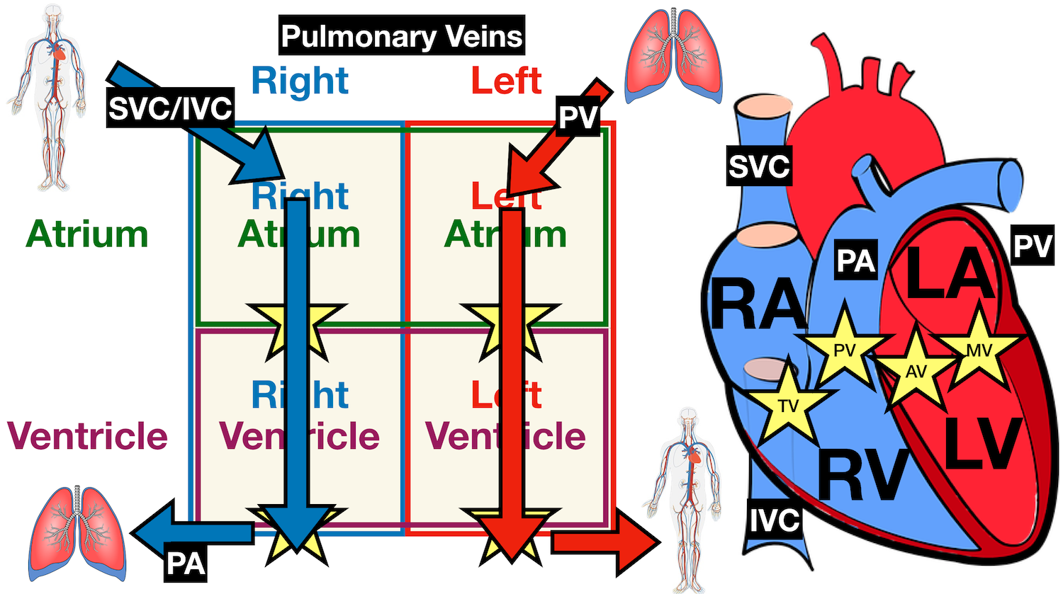
Image: Anatomy of the heart labeled diagram showing the main cardiac structures including the pulmonary veins.
Aorta
The final blood vessel is the aorta, and it is responsible for delivering oxygenated blood from the left side of the heart (left ventricle) to the rest of the body.
Again, we know the aorta is an artery as it is carrying blood away from the heart.
Image: Anatomy of the heart labeled diagram showing the main cardiac structures including the aorta.
Anatomy of the Heart
Now that we have reviewed the main anatomical structures of the heart using the cartoon image, let’s go back to that original diagram.
You should now be able to label all the main anatomical features.
Below is a blank diagram, followed by the labeled diagram with the answers.
Try to label the following structures yourself and then check to see if you are correct:
Right Atrium, Right Ventricle, Left Atrium, Left Ventricle, Tricuspid Valve, Mitral Valve, Pulmonary Valve, Aortic Valve, Superior Vena Cava, Inferior Vena Cava, Pulmonary Trunk, Right Pulmonary Artery, Left Pulmonary Artery, Pulmonary Veins, Aorta
Image: Diagram of the heart without labels to quiz yourself on the main cardiac structures and anatomy.
Now check to see if you got them all correct!
Image: Anatomy of the heart labeled diagram showing the main cardiac structures. (Not labeled: main pulmonary artery/pulmonary trunk emerging from the right ventricle and splitting into the right and left pulmonary arteries).
Before You Go….
Save yourself time, improve your studying, and help your career! Make sure to sign up for FREE to the EZmed blog down below, and don’t miss out on weekly updates on future medical and science topics made easy!
A weekly notification is sent right to your inbox filled with new blog posts, new videos, and exam prep!
Enjoy the post? Leave a comment down below!
Feedback or suggestions for future topics? Reach out using the contact button!
Thank you for using EZmed!
Make Your Learning Experience Even Easier!
Perform well in class, ace your exams, and keep up with your medical knowledge throughout your career using the following EZmed platforms:
YouTube Channel: EZmed - Animations and videos that simplify medicine and science
Instagram: @ezmedlearning - High yield exam content
Pinterest: ezmedlearning - Easy illustrations and flashcards